Home
/ Compact Bone Diagram Lacunae / Haversian Canal Wikipedia : Label compact and spongy bone illustrations as demonstrated in class.
Compact Bone Diagram Lacunae / Haversian Canal Wikipedia : Label compact and spongy bone illustrations as demonstrated in class.
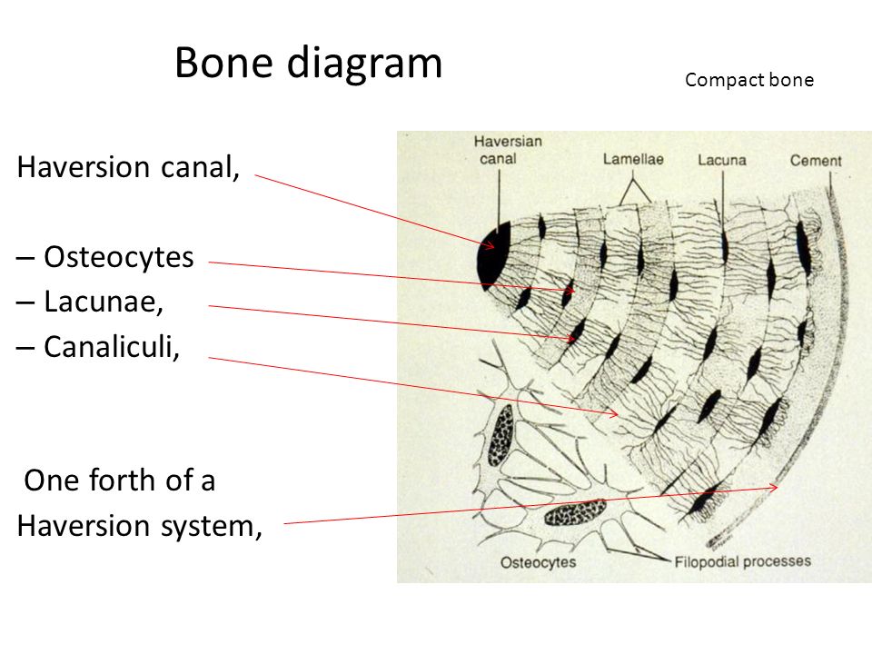
Compact Bone Diagram Lacunae / Haversian Canal Wikipedia : Label compact and spongy bone illustrations as demonstrated in class.. To know the structures of a synovial joint and a symphysis joint (intervertebral disc). Compact bone forms the surface of all bones. The osteon consists of a central canal called the osteonic (haversian) canal, which is surrounded by concentric rings (lamellae) of matrix. Compact bone, also called cortical bone, is the hard, stiff, smooth, thin, white bone tissue that surrounds all bones in the human body. They allow blood vessels and nerves to travel through them to supply the osteocytes.
The lacunae is the space that holds the cartilage in the bones of the human body. Lacunae possess canaliculi where each lacuna consist of only one cell (osteocyte). Compact bone is the denser stronger of the two types of bone tissue. The compact bones form the hard exterior of the bones, whereas the spongy bones have several pores that are filled with nerves and blood vessels. Osteon model lacunae canaliculi osteocyte.
Structure Of Compact Bone Longitudinal And Cross Sectional View Of Download Scientific Diagram from www.researchgate.net The compact bones form the hard exterior of the bones, whereas the spongy bones have several pores that are filled with nerves and blood vessels. Diagramme schnell und einfach erstellen. Compact bone is the denser, stronger of the two types of bone tissue ( (figure) ). Osteon model lacunae canaliculi osteocyte. The canaliculi give the osteon the appearance of having tiny cracks in the lamellae. Compact bone diagram microscope / bone structure anatomy and physiology i. Identify structures and functions of the microscopic structure of compact and spongy bone 3. The spaces between the trabeculae contain red or yellow marrow, depending on a person's age and on which bone it is.
Compact bone is the denser, stronger of the two types of bone tissue ( link ).
Compact bone diagram bone cross section diagram file624 diagram of compact bone new. Compact bone diagram microscope / bone structure anatomy and physiology i. Learn vocabulary, terms, and more with flashcards, games, and other study tools. Compact bone, also called cortical bone, dense bone in which the bony matrix is solidly filled with organic ground substance and inorganic salts, leaving only tiny spaces (lacunae) that contain the osteocytes, or bone cells.compact bone makes up 80 percent of the human skeleton; Between the rings of matrix the bone cells osteocytes are located in spaces called lacunae. The canaliculi give the osteon the appearance of having tiny cracks in the lamellae. Some, mostly older, compact bone is remodelled to form these haversian systems (or osteons).the osteocytes sit in their lacunae in concentric rings around a central haversian canal (which runs longitudinally).the osteocytes are arranged in concentric rings of bone matrix called lamellae (little plates), and their processes run in interconnecting canaliculi. It can be found under the periosteum and in the diaphyses of long bones, where it provides support and protection. Berapa harga sunscreen skin aqua. 3 mature bone cells, osteocytes, are found in tiny cavities within the matrix called lacunae. Osteocytes occupy spaces (lacunae) in the bone matrix. Compact bone is dense so that it can withstand compressive forces, while spongy. Lacunae possess canaliculi where each lacuna consist of only one cell (osteocyte).
Describe the process of bone formation and bone remodeling from fetus through adulthood compact bone • dense bone Compact bone diagram osteon compact bone ap pinterest anatomy human anatomy and. Compact bone, also called cortical bone, is the hard, stiff, smooth, thin, white bone tissue that surrounds all bones in the human body. The compact bones form the hard exterior of the bones, whereas the spongy bones have several pores that are filled with nerves and blood vessels. Compact bone accounts for 80% of the bones in the human body.
C Compact Bone Tissue A Canaliculi B Lacunae C Volkmann S Download Scientific Diagram from www.researchgate.net The spaces between the trabeculae contain red or yellow marrow, depending on a person's age and on which bone it is. In long bones, as you move from the outer cortical compact bone to the inner medullary cavity, the bone transitions to spongy bone. Compact bone, also called cortical bone, dense bone in which the bony matrix is solidly filled with organic ground substance and inorganic salts, leaving only tiny spaces (lacunae) that contain the osteocytes, or bone cells.compact bone makes up 80 percent of the human skeleton; Anatomy a cavity space or depression especially in a bone containing cartilage or bone cells. Haversian canals (sometimes canals of havers) are a series of microscopic tubes in the outermost region of bone called cortical bone. It can be found under the periosteum and in the diaphyses of long bones, where it provides support and protection. Diagramme schnell und einfach erstellen. It can be remodeled all throughout life to withstand stress.
Diagram of a typical long bone showing both cortical (compact) and cancellous (spongy) bone.
We discuss their function, the different types of bones in the human body, and the cells that are involved. Between the rings of matrix the bone cells osteocytes are located in spaces called lacunae. Label compact and spongy bone illustrations as demonstrated in class. How do nutrients reach the osteocytes in compact bone? What are your bones made of? Compact bone and spongy bone anatomy flashcards quizlet anatomy physiology exam 2. The compact bones form the hard exterior of the bones, whereas the spongy bones have several pores that are filled with nerves and blood vessels. Haversian canals (sometimes canals of havers) are a series of microscopic tubes in the outermost region of bone called cortical bone. The trabeculae are only a few cell layers thick. Compact bone diagram bone cross section diagram file624 diagram of compact bone new. Anatomy of a long bone proximal epiphysis diaphysis distal epiphysis compact bone spongy bone medullary cavity. Having been constructed in the 16th century, microscopes have revolutionalized science with their ability to magnify small objects such as microbial cells, producing images with definitive. Osteocytes, located in lacunae, are connected to one another by processes in canaliculi.
Compact bone diagram osteon compact bone ap pinterest anatomy human anatomy and. Compact bone consists of closely packed osteons or haversian systems. Compact bone, as opposed to spongy bone, is made of cylindrical units, called osteons, that are tightly formed together. Bone marrow is present (it is a kind of haematopoietic tissue from which all blood cells are made). Between the rings of matrix, the bone cells (osteocytes) are located in spaces called lacunae.
Medical School Histology Basics Cartilage And Bone Ppt Video Online Download from slideplayer.com Between the rings of matrix, the bone cells (osteocytes) are located in spaces called lacunae. Compact bone accounts for 80% of the bones in the human body. Compact bone consists of closely packed osteons or haversian systems. Compact bone diagram osteon compact bone ap pinterest anatomy human anatomy and. Identify structures and functions of the microscopic structure of compact and spongy bone 3. Some, mostly older, compact bone is remodelled to form these haversian systems (or osteons).the osteocytes sit in their lacunae in concentric rings around a central haversian canal (which runs longitudinally).the osteocytes are arranged in concentric rings of bone matrix called lamellae (little plates), and their processes run in interconnecting canaliculi. They allow blood vessels and nerves to travel through them to supply the osteocytes. Having been constructed in the 16th century, microscopes have revolutionalized science with their ability to magnify small objects such as microbial cells, producing images with definitive.
Of bone figure 5.3 page 116 objectives 1.
We discuss their function, the different types of bones in the human body, and the cells that are involved. The osteon consists of a central canal called the osteonic (haversian) canal, which is surrounded by concentric rings (lamellae) of matrix. Compact bone, as opposed to spongy bone, is made of cylindrical units, called osteons, that are tightly formed together. Compact bone diagram bone cross section diagram file624 diagram of compact bone new. A structural unit of compact bone consisting central haversian canal. It can be found under the periosteum and in the diaphyses of long bones, where it provides support and protection. The spaces between the trabeculae contain red or yellow marrow, depending on a person's age and on which bone it is. Anatomy of a long bone proximal epiphysis diaphysis distal epiphysis compact bone spongy bone medullary cavity. Compact bone is the denser stronger of the two types of bone tissue. The marrow in these images is red marrow. Canals between the lacunae of ossified bone. The trabeculae are only a few cell layers thick. Compact bone consists of closely packed osteons or haversian systems.

